
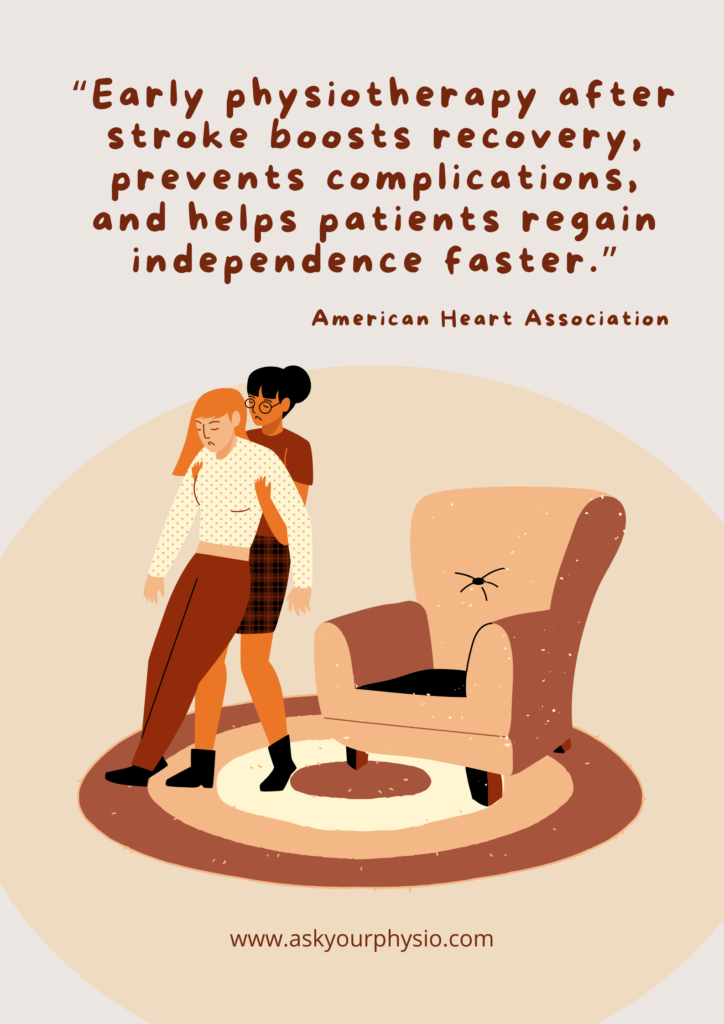
The stroke physical therapy is more productive & compelling when started earlier along with high intensity of exertion. Studies on stroke recovery have shown that the brain has the tendency to recuperate from the state of shock when the penumbra is stimulated timely. This can be achieved through the phenomenon of neuroplasticity that is the brain’s ability to rewire & adapt to new functions. When we say Stroke Rehabilitation: a complete physiotherapy guide to assessment, diagnosis & recovery, we mean thoroughgoing facts about it.
Table of Contents
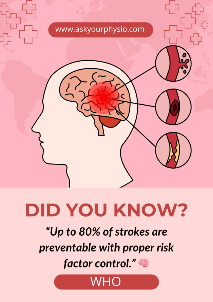
ToggleAccording to research from the World Health Organization and the American Heart Association, commencing physiotherapy during the initial 24 to 48 hours following a neurologically controlled stroke can greatly enhance the functional results. Mobility, speech, and routine activities can be restored within this time frame by incorporating the motor relearning techniques mainly task-specific and repetitive task training by assisting the higher brain centers to create new neural pathways.
The Clinical Manifestations of Stroke
Motor
- Paralysis
- Weakness
- Muscle Atrophy
- Gait Impairment
- Balance Impairment
Sensory
- Dermatomes
- Sensory Aphasia
- Pain
- Loss of sensations i.e, Temperature, Vibration, Light & Crude touch
- Proprioception
- Hemianopsias
- Neglect Syndrome
Cognitive
- Perceptual Disorders
- Loss of Executive Functioning
- Loss of Orientation
- Memory deficits
- Prosopagnosia
- Agnosia
Autonomic
- Autonomic Dysreflexia
- Loss of Fight & Flight Function
Post Stroke Pain Syndrome
It is characterized as a set of chronic disorders, especially the pain conditions that may occur after stroke because of harm to the parts of the brain that process all the sensory data. One of the most common types is the Central Post Stroke Pain, that arises when the lesions begin to affect the sensory networks such as the spinothalamic tracts or their relay center thalamus. Patients report a feeling of burning, electric current or shock, stabbing like pain frequently with abnormal sensations such as sensitivity to temperature & touch. According to research, 8 – 12% of stroke patients experience this kind of neuropathic discomfort weeks following the first incident.
The Types of Stroke
Stroke can be Ischemic or Hemorrhagic & with that it can be characterized into further types depending on the site of lesion.
Middle Cerebral Artery Stroke
- Characterized as the contralateral motor deficit
- Arms & Face are more affected as compared to Lower limb
- MCA stroke affecting the dominant hemisphere leads to aphasia
- MCA stroke affecting the non dominant hemisphere leads to impairments related to vision such as neglect syndrome, homonymous hemianopsia
- About 90 to 95% of people have left dominant hemisphere, yet in order to rule out which hemisphere is exactly dominant we need to look into the Edinburgh Handedness Inventory
Edinburgh Handedness Inventory
It comprises of 20 items:
- Writing
- Drawing
- Throwing
- Scissors use
- Combing
- Toothbrush use
- Knife without fork
- Spoon
- Hammer
- Screwdriver
- Tennis racket
- Knife with fork
- Cricket bat
- Golf club
- Broom
- Rake
- Striking a match
- Opening a box lid
- Dealing with cards
- Threading a needle
For each item, the participant is requested to choose which side they desire to complete the tasks with (self-assessed method); or the participants are required to complete the task and the results are evaluated by the therapist (direct observation technique).
Anterior Cerebral Artery Stroke
- Characterized as the contralateral motor deficit
- Lower limb is more affected as compared to upper limb & face
- Balance is the most compromised in these types of strokes
- The Frontal lobe is engaged so personality disorders such as agitation, behavior changes are more evident
- Appearance of Frontal Release Signs i.e, the re-appearance of the primitive reflexes as a result of destruction of inhibitory control of the frontal lobe & are suppressed as the frontal lobe matures with age. Common frontal release signs include Grasp reflex, Snout, Palmomental, Rooting, Sucking reflex, Glabellar etc. These can be assessed using Frontal Assessment Battery tool
Frontal Assessment Battery (FAB)
It is a brief bedside tool used to examine the executive functioning & frontal lobe functioning. It comprises 6 mini tasks that are to be performed each focuses on a distinct facet of executive function, and in a clinical environment, the entire evaluation typically takes five to ten minutes to do. It consists of 6 sets of 0 to 3 score with a maximum of 18. The higher the score, the better the frontal lobe is functioning.
- Conceptualization of Similarities
- Mental flexibility with verbal expression
- Motor tasking
- Interference sensitivity creating conflicts
- Inhibitory control a Yes/No/Yes situation
- Prehension behavior & autonomy of actions
Posterior Cerebral Artery Stroke
- Motor paralysis is not evident in this type of lesion; more sensory deficits
- Individual can walk & perform motor tasks with least alterations & is almost near to normal
- More affect to visual aspects & often called “Visual Strokes” causing issues such as diplopia, homonymous hemianopsia, visual agnosias etc
- If dominant hemisphere is involved then patient may exhibit signs of Alexia without the presence of Agraphia indicating that he can write but cannot read
Cross Deficits Following a Stroke
Cross deficits are the impairments that occur on the contralateral side to the lesion in the brain. This occurs as a result of the decussation of the motor & sensory tracts in the brainstem’s medulla, causing lesion of one hemisphere to involve the opposite side of the body.
Diagnosing the Stroke
- The faster the detection, the better the outcome – Rapid bedside examinations are often the first step in the early detection of stroke. The Face drooping, Arm weakness, Speech difficulty & Time to call help is the most extensive tool used globally. It assists medical professionals and the general people in promptly identifying potential stroke symptoms and starting critical care.
- The NIH scale to detect the extent of deficit – the gold-standard diagnostic instrument for measuring the degree of severity of a stroke is the NIH scale. Consciousness, gaze, vision, face movements, limb strength, sensations, speech, and neglect are all assessed. More severe neurological damage is indicated by higher scores, which range from 0 to 42.
- CT or MRI help – Neuroimaging, such as a computed tomography scan or magnetic resonance imaging, is necessary for an accurate evaluation. Since a CT scan can swiftly distinguish between an ischemic stroke and a hemorrhagic stroke, a crucial step in determining the appropriate course of treatment, it is typically done promptly.
- Functional Assessment Scales to plan the road to recovery – once the diagnosis is confirmed, the functional scales can be used to assess the level of functionality. These include:
- The Barthel Index – used to evaluate the independence of activity levels with scores ranging from 0 to 100
- Modified Rankin Scale – assesses the overall extent of disability following a stroke with score 0 which indicates no symptoms to 6 meaning death
- Functional Independence Measure – to evaluate the overall psychological, physical & social functioning
- Stroke Impact Scale – a tool utilized to assess the strength, grasping or hand function, memory, cognition, ADLs & participation. This scale tells about the overall quality & standard of life
- Rivermead Mobility Index – for mobility & function specifically
The Recovery Window Intervention Exercises
The goal of therapy during the first six months following a stroke is to promote healing with monotonous, task-specific exercises that promote neuroplasticity. Since this phase is thought to be the most sensitive for motor relearning, physiotherapy places a strong emphasis on regaining independence in daily activities, durability, movement and synchronization.
Evidence based Information
When compared to delayed rehabilitation, a clinical investigation on stroke patients indicated that beginning therapy within 24 hours greatly improved neurological results. Patients who began therapy early showed significantly higher recovery scores after three months of rehabilitation. The results showed that
- The early-rehab group’s NIHSS score increased from 3.21 to 0.76.
- In comparison to the deferred rehabilitation category, motor function as assessed through the Fugl-Meyer Assessment improved to 91.18 points after three months.
- Exercise capacity, daily tasks, and overall quality of life all showed improvements.
List of exercises to be Implemented as Intervention:
- Patient & care takers’s counseling
- Range of motion exercises
- Bed mobility & transfers
- Strengthening exercises
- Balance & Posture
- Gait training
- Task oriented training
- Upper limb training CIMT
- For more details read our previous blog 8 Essential Facts to know about Stroke Rehab Recovery
This article has been written by a Physical Therapist and provides general guidance on physical health & exercise. While it is grounded in professional expertise, it is not a substitute for individualized medical advice. If you are experiencing pain, specific symptoms, or have an underlying medical condition, please book a 1 on 1, 30 minute consultation with our expert physical therapist for a personalized assessment & tailored recommendations.